
VINNO 8 brings premium performance without bounds. It combines advanced zone imaging technology, smart workflow, compact and light weight design all in one.
Advanced Imaging Technologies
Enhanced OB/GYN Application
Extended Cardiac Application
Innovative Design
Discover More

Advanced Imaging Technologies
Zone Imaging
Zone Imaging technology acquires better image resolution and energy distribution in entire zone area through apodizing transmission in multiple frequency and instantaneous phase superposition.

Transmit
Apodizing transmission in Multiple Frequency
Focus
Focus in entire zone area through instantaneous phase superposition
Receive
Compound in group phases via coherence technique
Imaging
Acquire the images after grid cross processing
VFlow
VFlow is an advanced adaptive filter technology of color flow from VINNO to enhance the detective sensitivity of tiny blood flow.


Pure Wave Probe Technology
Better orientation delivers better penetration to difficult person.
Advanced Imaging Technologies

Volume Contrast Imaging (VCI)
Adjust slice thickness on 2D planes in rendering 3D or 4D mode to enhance contrast resolution.

Niche View
Reconstruct a volume structure with multiplanar views, better reveal the spatial correlation of tissue structure.

Multiline Free View
Obtain any plane from a volume data by simply drawing a line, curve, through a structure. This valuable technology enables views of even irregularly shaped structures not attainable in 2D imaging. Now we provide up to 3 Free view images displayed simultaneously.

Hysterosalpingo Contrast Sonography (HSG)
A simple and well-tolerated ultrasound procedure used to access the patency of the fallopian tubes and the abnormalities of the uterus and endometrium.
Ergonomically Designed for Daily Use
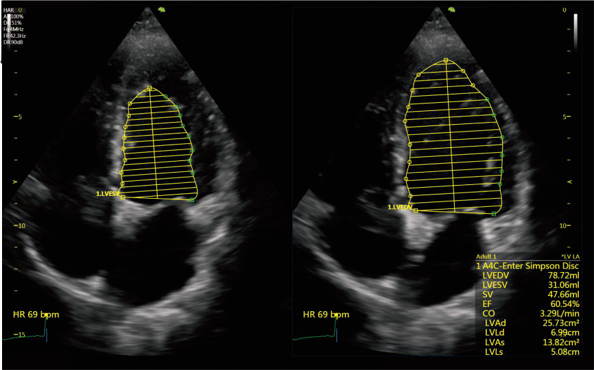
Auto EF Measurement
The system can calculate cardiac function of EF values through clicking three
different key positions instead of tracing the whole endocardium edge by manual,
which saves a lot of time.
Various Imaging Modes for Accurate Cardiac Analysis

CW

TDI

Multi-Angle M Mode (MAM)

Stress Echo
Discover More
Simple To Learn
Intuitive touch panel operation and built-in user manual for reference
Precise Imaging
2-23Mhz frequency range enables you see from superficial to deep tissue
Easy Compare
Before and after exam comparison with Easy Compare function
Accurate Biopsy Guidance
Needle enhancement makes the needle tip more visible
Hysterosalpingo Contrast Sonography (HSG)
Easy Compare for patient exam follow up.
Needle Enhancement is a nice tool to visualize the needle tip in radiology interventional application.
Contrast Bubble Imaging is good for organ edge delineation; monitoring blood perfusion in organs and recognition of lesion characterization.


